Blog description

For students to fill in

Thursday, 4 December 2014

Hormonelle Veränderungen / Sarah


Hormonelle Veränderungen während der Pubertät


Hormone sind chemische Stoffe, die die Körpervorgänge steuern, wie das Wachstum, den Stoffwechsel.. Sie entstehen in den Hormondrüsen.

Die Pubertät beginnt bei den Mädchen zwischen dem zehnten und dem 13. Lebensjahr, und bei den Jungen erst zwischen dem 12. und dem 15. Lebensjahr

Wenn der Körper bereit ist, geschlechtsreif zu werden, sendet der Hypothalamus, ein Teil des Zwischenhirns, chemische Signalstoffe zur Hypophyse, die an der Unterseite der Gehirns liegt. 


Die Hirnanhangdrüse und der Hypothalamus sind durch den so genannten Hypophysenstiel verbunden, auf diese Weise erhält die Hirnanhangdrüse  genaue Informationen über die körperliche Entwicklung und produziert abhängig davon die richtige Anzahl an Hormonen. 


Die Hypophyse besteht aus drei Teilen, dem Hypophysenvorderlappen, dem Hypophysenhinterlappen und dem Hypophysenzwischenlappen.

In dem Hypophysenvorderlappen werden die folgenden Hormone produziert :

-Somatropin, was für das Körperwachstum zuständig ist. Diese Hormone werden meistens während des Schlafes produziert, und sie sind essentiell für ein normales Wachstum. 

-Follikelstimulierendes Hormon (FSH): Es bewirkt das Wachstum von Samenzellen bei Männern und der Eizellen in den Eierstöcken bei den Frauen. 

-Luteinisierendes Hormon (LH):  Es regt bei der Frau den Eisprung und beim Mann die Reifung der Spermien an.

-Östrogen: Sie sind die wichtigsten weiblichen Sexualhormone. Sie werden hauptsächlich in den Eierstöcken produziert. Sie steuern die Vorgänge des weiblichen Zyklus' und der Fortpflanzung und sorgen bei Mädchen für das Wachstum der Geschlechtsorgane und der Ausbildung der Schleimhaut. Sie sind auch dafür zuständig, dass während der Pubertät die Brüste wachsen und eine rundliche Körperform entsteht.

-Testosteron: Ist das wichtigste Sexualhormon bei den Männern. Es wird vor allem in den Hoden gebildet unter dem Einfluss von LH. Es fördert das Wachstum und die Entwicklung der Geschlechtsorgane. Es ist für die männlichen Geschlechtsmerkmale wie eine tiefe Stimme, Bartwuchs, breiten Schultern oder Muskelaufbau verantwortlich. Es wirkt auch auf den Sexualtrieb und auf die Samenproduktion.


meine Quellen: 

http://www.helles-koepfchen.de/artikel/3404.html


http://de.wikipedia.org/wiki/Pubertät


Sunday, 30 November 2014

Diabetes and Oxytocin // Katrina Dombovári // S6EN

In the last two weeks, we have been learning about type one and type two diabetes, and a bit about oxytocin. 

Starting off with diabetes, type one diabetes is the worst form of diabetes. Type one diabetes effects mainly younger people. When someone has type one diabetes it means that the beta cells are destroyed and the levels of glucose in the person’s blood is too high. Someone with type one diabetes needs to have insulin injected in them every so often (around twice a day), to insure that the glucose levels don’t get too high. Someone with type one diabetes also needs to watch their diet closely. 

The other form of diabetes is type two (diabetes). Type two diabetes mainly affects overweight and older people. Someone with type two diabetes has an impaired insulin secretion, insulin resistance and their level of glucose in the blood is too high. What that means is that the body (pancreas) of someone with type two diabetes can make insulin properly (unlike someone with type one diabetes), however either that person’s pancreas doesn’t make enough insulin to keep the glucose level stable, or the body can’t use the insulin well enough. Just like people with type one diabetes, people with type two also need to monitor their diet closely. 

For a long while, there wasn’t any form of treatment for type one diabetes. However around the 20th century, a Canadian scientist, by the name of Frederick Banting, decided that he would remove the pancreas of a dog, since that’s where diabetes is effected, and see what happens. To do this he needed financial support, which he got, by a Canadian biochemist, McCloud. Banting was also assisted by Best, a scientist.

After they extracted the pancreas from the dog, the blood sugar of the dog rose, the dog became more thirsty, it drank a lot of water, urinated more often, and all in all, the dog became much more weaker. In conclusion, they found out that the dog had developed diabetes. The scientists managed to save the dog though! To save the dog, the scientists injected a healthy version of the extracted pancreas (also known as insulin). This causes the previous effects on the dog to be reversed, meaning it became stronger, the blood sugar stayed level and it wasn’t as thirsty as it was before. To keep the dog healthy, and to insure that the previous effects wouldn’t return, they needed to continue to inject the dog with insulin everyday. 

Banting and McCloud then won the noble prize to figuring out a treatment for type one diabetes. 

In class we had to do a article about the history of diabetes. This is my article:

History of Diabetes

Diabetes mellitus was found around the Egyptian time. For a long period of time it was known as a disease of the kidneys from around the 19th century. It was recognized by it's excessive amount of urine output. Observations were made by Areteus of Cappadocia, which show that it effects the kidneys. Someone took their urine and tasted it, which helped them tell if they had type one diabetes or not. This person who tested their urine in 1674, recognized that the urine had a sweet taste to it, which is how diabetes mellitus got it's name (diabetes meaning 'to run through'; mellitus meaning 'sweet like honey'). The sweet taste of the urine was caused because of the high glucose levels in the blood. 

For a long period of time no one was able to find a treatment. In 1869 a German medical student, Paul Langerhans, found out that in the tissues of the pancreas produces digestive juices in a cell with an unknown function. These cells were found to be the beta cells, which produce insulin. 

In 1923, Banting, a Canadian scientist, decide to try to extract the pancreas of a dog to see what would happen to that dog- would this dog get diabetes or not? Banting was supported by another Canadian biochemist, McCloud, and was assisted by a scientist by the last name of Best.

What they did was extract the pancreas of a dog. The removal of the pancreas caused it's blood sugar to rise and caused the dog to become weaker and weaker. Due to the removal of the pancreas, the dog got diabetes. They continued to try this on other dogs, and the same thing happened. What they then did was sliced up the pancreas into pieces and froze all the pieces in water and salt. After the pieces were half frozen, they were filtered, which then produced 'isletin'. The extract was injected back into a diabetic dog, and they observed that the blood glucose levels dropped and the dog became healthier once again. They gave the diabetic dog injections everyday to keep its blood glucose level stable, and the dog remained healthy and happy. 

In 1922, a 14 year old Canadian boy by the name of Leonard Thompson, was the first human being tested on with the insulin. Luckily, the test was very successful.

Due to the success of finding out how to treat type one diabetes, Banting and McCloud were rewarded a noble prize in Medicine and Physiology in 1923. 


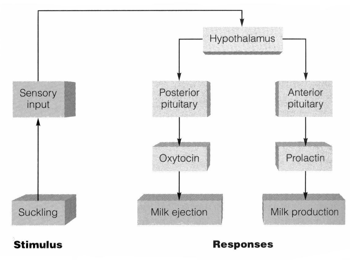
After learning about diabetes, we began to talk a bit about oxytocin. Oxytocin is a hormone, that is made in the brain, in the hypothalamus and it gets transported to the pituitary gland. The release of oxytocin acts to regulate two female reproductive functions: childbirth and breast-feeding. Oxytocin is an important hormone for milk ejection. Its release is stimulated by seeing or hearing a stimulus, for example, the most intense release is triggered by the baby suckling on the mother’s breast. The first release is within a minute of suckling and continuously releases in spurts. It peaks and levels off about every six to ten minutes after nipple stimulation has stopped.


Sources of images:

 http://www.mchip.net/sites/default/files/mchipfiles/Oxytocin%20reflex.JPG

http://projects.cbe.ab.ca/Diefenbaker/Biology/Bio%20Website%20Final/notes/reproductive/Image72.gif

- Katrina Dombovári 

Wednesday, 19 November 2014

Wirkung von Alkohol auf unser Nervensystem / Anna Biermann S6DE


Wirkung von Alkohol auf unser Nervensystem

Der Konsum von Alkohol hat Einfluss auf unser Verhalten: schon nach kleinen Mengen fühlen wir uns selbstbewusster, haben verstärkten Tatendrang, reden mehr, bekommen Glücksgefühle. Je mehr Alkohol man zu sich nimmt desto stärker sind allerdings die Auswirkungen: Gleichgewichtsstörung, Verlangsamung der Reaktionsfähigkeit, Probleme beim Sprechen bis hin zur Bewusstlosigkeit.

Das heißt also, dass die Reizübertragung durch das zentrale Nervensystem unter dem Einfluss von Alkohol nicht mehr gut funktioniert.

WIESO?

Alkohol gelangt über die Blutbahn in den gesamten Körper und ist in ca. 2 Minuten auf diesem Weg im Gehirn. In kleineren Mengen wirken alkoholische Getränke anregend, in größeren Mengen dagegen berauschend. Die dafür verantwortliche Substanz im Alkohol heißt Ethanol (C2H6O). Alkohol löst eine biochemische Kettenreaktion in unserem Körper aus.
Der Alkohol verteilt sich im ganzen Körper und beeinflusst alle Transmittersysteme im Nervensystem.

Neurotransmitter sind nichts anderes als Botenstoffe, die an den Synapsen Informationen zwischen den Nervenzellen übertragen.

https://www.youtube.com/watch?v=BEglM5JehY4


Glutamat, Gaba, Serotonin, Dopamin, Acetylcholin sind solche Neurotransmitter.   
Wenn nun Alkoholmoleküle sich an die Rezeptoren der Nervenzellen binden, verändern sie die Informationsübertragung zwischen den Nervenzellen. Das heißt, die Funktionsweise der Neurotransmitter wird gestört. 
Wenn wir uns schon nach einem Glas Wein enthemmter, lustiger, zufriedener fühlen, dann sind dafür die Neurotransmitter Serotonin und Dopamin verantwortlich, die sogenannten Glückshormone. Serotonin gilt als zentraler Stimmungsmacher und beeinflusst unter anderem Appetit, Sexualtrieb und das psychische Wohlbefinden. 
Dopamin ist für Aufmerksamkeit, Wohlbefinden, Lernfähigkeit und motorische Aktivitäten zuständig. Die von den Alkoholmolekülen angegriffenen Nervenzellen schütten nun vermehrt Serotonin und Dopamin aus, die vom Belohnungszentrum im Gehirn über spezielle Rezeptoren aufgenommen wird.


Die Transmittersysteme, Gaba und Glutamat sorgen für das Gleichgewicht der Gehirnaktivität. Glutamat ist für die Gehirnaktivität zuständig, für die Übermittlung von Sinneswahrnehmungen, Bewegungssteuerung sowie für das Lernen und das Gedächtnis. Im Gehirn ist Glutamat der häufigste erregende Neurotransmitter. Gaba ist der Transmitter für die Dämpfung der Gehirnaktivität und somit gewissermaßen der Gegenspieler von Glutamat. Gaba ist der wichtigste hemmende Neurotransmitter im Gehirn, also  der wichtigste hemmende Botenstoff von Nerv zu Nerv.

Der Alkohol bringt das Gleichgewicht von Gaba und Glutamate durcheinander. Alkohol hat eine dämpfende Wirkung auf das Gehirn. Der aktivierende Botenstoff Glutamat wird gedämpft und die hemmende Wirkung von Gaba durch den Alkohol verstärkt.

So erklären sich also die oben genannten Symptome wie Gleichgewichtsstörung, Verlangsamung der Reaktionsfähigkeit, Probleme beim Sprechen bis hin zur Bewusstlosigkeit in Folge von Alkoholkonsum.

Alkohol ist ein gefährliches Nervengift und kann süchtig machen.


Quellen:


Wednesday, 5 November 2014

Vaskuläre Demenz

VASKULÄRE DEMENZ


Heute möchte ich euch eine andere Form der Demenzerkrankung vorstellen. Die vaskuläre Demenz ist nach Alzheimer die verbreitetste Demenzkrankheit.


Was ist die VASKULÄRE DEMENZ?
Die vaskuläre Demenz zählt zu den Demenzkrankheiten. Oft kann es passieren, dass sie mit Alzheimer verwechselt wird. Dem ist aber nicht so, da diese Form der Demenz eine ganz andere Ursache hat. Man bezeichnet sie eben als vaskulär (vaskuläre=Blut/Blutgefäße), da sie sich aufgrund von Durchblutungsstörungen im Gehirn entwickelt. Die große Eigenart an dieser Krankheit sind ihre unterschiedlichen Symptome die nicht vorhersehbar sind. Wie Alzheimer auch ist die vaskuläre Demenz nicht heilbar.


                                                                      Ursachen:

Die Ursachen einer vaskulären Demenz sind auf Durchblutungsstörungen zurückzuführen. Sie lösen im Gehirn eine Kettenreaktion aus. Eine verengte Arterie wird durch eine kleine Kruste verstopft. Dadurch steigt der Druck in der Arterie und sie platzt. Jetzt werden manche Teile des Gehirns nicht mehr mit Sauerstoff versorgt und sie fangen langsam an abzusterben. Man nennt das auch einen Schlaganfall. Da unser ganzes Gehirn mit Arterien verbunden ist, ist es reiner Zufall welche Hirnregionen betroffen werden, denn entsprechend sind die Symptome jedes Mal etwas anders. Die Stärke der Erkrankung ist davon abhängig ob es ein großer oder mehrere kleine Hirnschläge waren. Gefährlicher sind oft viele kleine, da hier besonders viele Regionen betroffen sind.


Symptome:
Die meisten Patienten wirken verwirrt, weil es ihnen immer schwerer fällt aufmerksam zuzuhören, sich zu orientieren und zusammenhängend zu reden. Diese Symptome treten meist früher und heftiger auf als bei Alzheimer. Dennoch behält der Betroffene oft meist noch sehr viel länger sein Gedächtnis. Andere Symptome können Persönlichkeitsveränderungen, nachlassendes Urteilvermögen und Probleme beim Alltag sein z.B weiß man nicht mehr wie der Fernseher angehet usw…


Therapie:
Wie Alzheimer ist auch diese Krankheit nicht heilbar. In der Regel gilt, ist das Gehirn einmal geschädigt ist es nicht mehr rückgängig zu machen. Es ist aber trotzdem wichtig die Krankheit frühzeitig zuerkennen um dem Patienten ein besseres Wohlbefinden für die Zukunft zu ermöglichen und um den Verlauf der Krankheit zu verlangsamen. Man kann dieser Demenz Erkrankung aber vorbeugen in dem man sich gesund ernährt und viel Sport macht.   


QUELLEN:
-http://www.alzheimer.de/alzheimer/alzheimer/weiteredemenzformen/vaskulaeredemenz.html

Monday, 3 November 2014

Diseases of the Nervous System - Stefanie Matei S6EN

In week 41 in biology 2p we started learning about hormones.

The course started off with a song by Frank Sinatra, which, by looking at the lyrics, we decided was about a certain hormone called oxytocin.

So what is oxytocin? Oxytocin is one of the most interesting hormones, which is centered in the brain. It deals with maternal instincts, love impulses, orgasms, but also anxiety and social recognition. When there is a lack of oxytocin, the individual may go through stages of depression, but if oxytocin is in abundance, then the individual is considered to have a higher and sharper sense of instinct -- some would go as far as saying it can give them unnatural attributes. 

Now that we learned what oxytocin is, we can go ahead and start with the basics of hormones. 

What is a hormone? A hormone is a messenger molecule that is transported by the circulatory system to get to organs in order to regulate the person's behaviour. Hormones work slowly, traveling through your bloodstream. They affect many processes such as metabolism, sexual function, mood, growth and physical development, which is why during your teenage years hormones are most at work.

The endocrine system is made of glands and organs, and is where hormones are produced, stored and released. The glands are stimulated by the nervous system. 

Hormones can come from three different origins: peptide, amine or steroid. 

A steroid hormone is derived from cholesterol and deals with a lot of aspects, though mainly it regulates your metabolism and controls your sexual characteristics. Examples of steroid hormones are 

An amine hormone mostly deals with digestion, and is mostly found in the intestinal or pancreas tissues.

A peptide hormone is made of protein. An example of a peptide hormone is actually one we saw earlier, which is oxytocin.


In week 42 each student of the course presented a disease of their choice that affected the brain or the nervous system. Here is the list:


Alzheimer's Disease

Alzheimer's is the most common version of dementia and usually comes with old age, patients diagnosed with it almost always being over 65 (though there are rare cases for younger patients). Alzheimer's is a disease that causes memory dissipation, starting out in stages of short term memory loss, then progresses into long term.

It was discovered in 1906 by Alois Alzheimer, a psychiatrist who first noticed it on one of his patients called Auguste D who had been suffering of it since 1901. It was made an official disease by Emile Kraepelin later on after more patients were discovered to have the same symptoms as Auguste D.

Usually, AD patients have a loss of function in the temporal lobe, the part of the brain that deals with memory. Here is a photo of how the brain deteriorates itself, to the left being a healthy brain, and to the right a case of alzheimer's:


There has so far been no discovered cure.


Mad Cow Disease

Bovine Spongiform Encephalopathy, otherwise known as mad cow disease is a neurodegenerative disease that can be caught by eating cow meat. It was named Mad cow disease because when cows got infected with it, their behavior changed, and they went crazy -- or mad. The human version is called Creutzfeldt-Jakob disease, and it affects the humans brain.

This disease is not contagious, and can only be caught if the person eats cow meat that contains contaminated central nerve tissues. The disease can't be transmitted from human to human, only by eating contaminated meat.

The disease is fatal. Once a patient is infected with it, their lifespan shrinks to more or less 13 months. 

There was a big Mad cow outbreak in the UK back in 2009, when 177 were killed by it. Since then, the US developed a law that stated that all brain and spinal cattle and cow meat is forbidden from being sold.


Epilepsy

Epilepsy is a general disorder, most common between teenagers and elders. 

Epilepsy happens when the brain recieves too many light and color signals that flash too fast, then becomes confused and triggers seizures. 

Here is an example of something that would induce epilepsy (open at your own risk):


So far, there has been no cure for epilepsy, only drugs that make the seizures stop but cannot prevent new ones from happening in the future.


Congenital indifference to pain

Congenital indifference to pain is a rare disorder that consists of the individual being able to feel touch or temperature, but not any physical pain.

How this happens: when a normal person gets hurt, the pain receptors (nociceptors) transmit a message through the nervous system (peripheral nerve) to the brain in order to tell that you're hurt. Here is a photo oh how it works:


For patients suffering from congenital indifference, the problem isn't that the brain doesn't recognize the pain, it's that the nerve tissues are either weakened or non existent, meaning that in the brain never gets any pain message since there's nobody sending them. 

Congenital indifference to pain is not something contagious, it is actually very rare, and can only be transmitted if both parents have a copy of the specific chromosome, making it a genetic disease.


Broca's aphasia

Broca's aphasia, otherwise known as expression aphasia, is a disorder that is characterized by the loss of being able to talk.

The most common cause of this disorder is a stroke, which is caused by a lack of brain oxygenation. The part of the brain usually affected and damaged is the bronca, hence the name of this aphasia. 

Brian's aphasia cannot be fully cured, but can become better with speech therapy and other expressive activities depending on how severe the case is.

Here is a video of how patients with Broca's aphasia speak:


Monday, 20 October 2014

The Scale of the Universe


Check out this amazing interactive infographic on the scale of the universe. What is even more incredible is that it was created by a pair of 14 year old twins!

Monday, 6 October 2014

Neurons and Neurotransmitters - Gador Ramos S6EN


         This past week we have been learning about neurons; their different parts and how do they interact. To understand better how neurotransmitter work, we had to choose an inhibitory or excitatory neurotransmitter and draw a comic about the effect the neurotransmitter has on people and how it works inside the brain.
         There are neurons all over our brain, they are minuscule and their function is to pass the information (electrical energy) from one neuron to another. The neuron that is passing the energy is called the presynaptic neuron and the neuron which is receiving the energy is the post synaptic neuron. The neuron is composed of different parts, the above part, which is a round shape type connected with thin lines (the dendrites), is called the cell body or the soma. Inside the soma there is the nucleus. There is a thicker line which is attached to the bottom of the cell body and it is called the axon. The axon is longer and thicker than the dendrites, and it is covered by myelin. Myelin is an insulating layer that prevents the electrical signal passing through the axon to escape from it. Then, the axon separates into different branches, and these branches separate into tinnier branches which are the axon terminals. At the end of the axon terminals is where the presynaptic terminals are located. Between the presynaptic terminal of the presynaptic neuron and the dendrites of the postsynaptic neuron, there is a space which is called the synaptic space or synaptic clef. The nerve impulse always goes to the right.

         In the presynaptic terminal all the energy is passed from one neuron to another. In the terminal there are vesicles filled in with neurotransmitters, when the energy comes to the terminal the vesicles go to the membrane and there, they free the neurotransmitters. 


The neurotransmitters being freed into the synaptic space go to their  receptors (attached to the dendrite) and pass the energy to the post synaptic neuron. Once they do that, all the receptors (the ones that have found their adequate receptors and the ones that have not) return to the presynaptic neuron passing through transporters.  
         When the axon is at rest and no electrical transmitters are passing through it, it has a negative charge on the inside and, on the outside it has a positive charge.  When electrical transmitters pass through parts of the axon it makes that part turn from a positive charge to a negative charge or vice versa.
         There are different neurotransmitters: neutral, excitatory and inhibitory ones.
These pass differently through the neurons. The neutral neurotransmitters make the electricity go at a normal rate, the excitatory neurotransmitters (ex: Glutamate) make the electricity go much faster than it should and lastly the inhibitory neurotransmitters make the electricity go at a very slow rate.
        
         To understand better how the excitatory or the inhibitory neurotransmitters work in real life, we had to draw a comic of how it worked inside our bodies but also on the outside and in what situations we use it or how it could help us.
I did the excitatory neurotransmitter norepinephrine. This neurotransmitter is like adrenaline, it comes from the medulla in the kidneys and it is responsible for stimulatory process in the body. It works by narrowing the blood vessels and increasing blood pressure and blood glucose levels. This is the process of my working comic and others working comics:


Sources of images: 1. vv.carleton.ca
                                2. cla.calpoly.edu


By Gádor Ramos S6EN

Tuesday, 30 September 2014

Biology of the Brain - Laura Darcy S6EN

This blog entry is about one of the most fascinating subjects in biology, the human brain. The supreme organ in the human body, the brain is what makes you, you. In the S6EN class we talked about the different functions of the brain and gave the example of a person cycling. A cyclist will be using various functions of the brain, and these are the things we listed in class:

  • the beating of the heart
  • physical movement
  • thinking
  • breathing
  • sweating
  • digesting
  • reading
  • perception
As you can see your brain has multiple and vital functions. Some of them are ordered by you and others are done without you even noticing. For each different function your brain has an area that deals with that specific job. For example what part of your brain are you using to read this right now? Well in fact it is not just one specific part but multiple parts of the brain that help you see, read and understand what I'm writing.
This brings us to what we did next. We were given a diagram similar to this one so that we would be more familiar with the names, functions and placement of things in the brain.

This helped us with the exercise we were given next. These are PET (positron emission tomography) images of the brain doing different activities. Using our knowledge of the brain, based on the diagram above, we were able to identify the activity in question.

One of the other things we talked about was the nervous system, although we didn't go into much detail. We learned that the nervous system is made up of two main groups, one called CNS (central nervous system) which includes the brain
and the spinal cord, amd the other one called PNS (periphiral nervous system)
which is made up of the nerves around the body.


Dissection of a brain

In this experiment, we dissected a calf's brain. We started by placing the brain in the tray. Then, with the available tools, we explored the different parts of the brain and tried to label them. I cut through a lot of parts in the brain to see the internal structure and I found out that they all had a similar structure on the inside. The brain was softer than I thought and it made it harder for me to handle. The dissection helped me understand how things were placed and at the same time helped memorize the different functions of specific areas in the brain.

the materials we used in this experiment were the following:

  • a tray
  • a straight needle
  • scissors
  • a scalpel
  • gloves
  • a lab coat
I positioned the brain in a way that allowed me to see the superior part of it. Although the two hemispheres detached from each other the moment I put the brain on my tray, the cerebellum was still firmily attached. This in a way made it easier for me to observe the two sides of the brain seperatly. The corpus callosum is the white stuff that holds the two sides of the brain together but in my case it was cut. The brain is filled with crevasses. The actual crevasse is called sulcus and the space between them is called gyrus.


Then cut open the cerebellum to examine the insides and saw the white and gray matter. 


I also cut the medulla open which was tougher and saw the beginning of the spinal cord. The picture also shows the pons on the right bottom corner.

I took the left side of the brain and cut it open and saw the same two kinds of matter that were in the cerebellum, the white and gray matter.

I observed and labelled different parts in the brain all throughout the dissection. I noticed how flexible and fragile the brain was at some parts. This made it harder for me to handle and when I cut one of the hemispheres of the brain open, the white matter seemed to almost fall out completely because of how soft it was. This is why the brain has maximum protection around it, like the skull, the cerebrospinal fluid that surrounds the brain to cushion it, and a series of membranes the first one being dura mater which acts as a kind of seatbelt. 

Obviously these were not on the brain when they were given to us otherwise it would have being a lot harder to cut through it. When cutting through different parts of the brain I realised that they were all composed of the same thing, grey and white matter. The white matter serves as a sort of insulator in order to keep the current flowing through the grey matter which is full of nerves. This helps avoid any chemical and electrical signals from neurons to move randomly, and ensures they reach the correct destination. I also learned that the reason why the brain has many folds (filled with veins) is to take less space and fit inside our skull.
What made this experiment difficult was labelling the different parts of the brain since there was no line telling you when it started and when it ended. I also never found the hippocampus.

 The brain of a calf is very similar to that of a human and so gives us a clear idea of what our brain looks like. It also helps us understand how the parts fit in a 3D space rather than looking at it on a flat piece of paper. After this dissection I am even more fascinated about the brain and the functions it performs. The brain is one of the most amazing organs, it keeps our heart beating without conscious effort and is parallel processing all the time. The brain is a highly complex organ that remains an object of scientific study.

References:
http://www.austincc.edu/apreview/PhysiologicalSystem/Nervous.html
http://www.dreamlanguage.org/articles/cognitive_function_in_dreams.htm

Thursday, 25 September 2014

Die Alzheimer Krankeit / Rachel

-Die Alzheimer Krankeit-


Alzheimer wurde von dem deutschen Nervenarzt Alois Alzeheimer entdeckt. 

Wir haben im Gehirn Milliarden von Nervenzellen (Neuronen), bei Alzheimer-Kranken sterben diese Nervenzellen langsam ab.
https://www.youtube.com/watch?v=0J8GY5yWqiI


Forscher haben noch kein Heilmittel gefunden, das heißt dass diese Krankheit bis jetzt leider unheilbar ist.


Die Alzheimer Krankheit hat drei verschiedene Stadien :

Im ersten Stadium hat der Patient  Orientierungsprobleme, Sprachschwierigkeiten und leichte Vergesslichkeiten.  Diese Phase ist kaum spürbar, man vergisst zum Beispiel seine eigene Telefonnummer oder man verwechselt Namen.

Später kommen andere Krankheitszeichen dazu die allerdings  problematischer sind. Die Person erkennt seine Familie, Freunde und Umgebung nicht mehr. Sie kann sich nicht sauber halten ; kochen und essen kann sie alleine auch nicht.
In dieser Phase braucht der Alzheimer Kranke ständig eine Person die ihm hilft. 

Dann beginnt das dritte Stadium. Diese Phase kann bis sieben Jahre dauern. Der Betroffene kann nicht mehr klar denken, kann sich kaum noch bewegen, hat Schwierigkeiten zu kauen und zu schlucken. In dieser Zeit kann der Alzheimer-Kranke sein Bett nicht verlassen.

Mehr Informationen: 

Alzheimer ist eine von den Krankheiten die im Alter auftreten.

http://www.alzheimerandyou.de/welcome/wie-haeufig-kommt-das-vor/

Weltweit leiden 20 Millionen Menschen an der Krankheit. In Deutschland sind es 1.5 Millionen. Diese Zahl wird sich bis 2050 bestimmt verdoppeln.

http://www.alzheimerandyou.de/welcome/wie-haeufig-kommt-das-vor/


http://www.google.fr/imgres?imgurl=http%3A%2F%2Fp7.storage.canalblog.com%2F73%2F19%2F562695%2F35015130.jpg&imgrefurl=http%3A%2F%2Falzheimer2.canalblog.com%2Farchives%2F2009%2F01%2F22%2F12184320.html&h=397&w=350&tbnid=L6rrmHA4mUYsTM%3A&zoom=1&docid=ILWAfuamIxpkdM&ei=1foiVKXSB7GLsQTcmILYDg&tbm=isch&iact=rc&uact=3&dur=613&page=1&start=0&ndsp=27&ved=0CCcQrQMwAA


Meine Quellen:

http://www.alzheimerandyou.de/welcome/wie-haeufig-kommt-das-vor/

http://www.wasistwas.de/wissenschaft/die-themen/artikel/link//11111/article/welt-alzheimer-tag-2003.html